
Steinberg Staging Of Avascular Necrosis/ Osteonecrosis
History and etymology
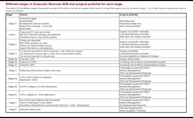
It is based on the radiographic appearance and location of lesion. It primarily differs from the other systems by quantifying the involvement of femoral head which allows direct comparison between series1. Seven stages of involvement are identified. Following staging, extent of involvement of femoral head is recorded as mild, moderate or severe.
Classification
stage 0: normal or non-diagnostic radiographs, MRI and bone scan of at risk hip (often contralateral hip involved, or patient has risk factors and hip pain)
stage I: normal radiograph, abnormal bone scan and/or MRI
stage II: cystic and sclerotic radiographic changes
stage III: subchondral lucency or crescent sign
stage IV: flattening of femoral head, with depression graded into
mild: <2 mm
moderate: 2-4 mm
severe: >4 mm
stage V: joint space narrowing with or without acetabular involvement
stage VI: advanced degenerative changes
Quantification of extent of involvement is necessary for stages I to V:
stage I and II
A, mild: <15% head involvement as seen on radiograph or MRI
B, moderate: 15% to 30%
C, severe: >30%
stage III
A, mild: subchondral collapse (crescent) beneath <15% of articular surface
B, moderate: crescent beneath 15% to 30%
C, severe: crescent beneath >30%
stage IV
A, mild: <15% of surface has collapsed and depression is <2mm
B, moderate: 15% to 30% collapsed or 2 to 4mm depression
C, severe: >30% collapsed or >4mm depression
stage V
A, B or C: average of femoral head involvement, as determined in stage IV, and estimated acetabular involvement.
Steinberg Staging Osteonecrosis
See also
References
1. Steinberg ME, Hayken GD, Steinberg DR. A quantitative system for staging avascular necrosis. J Bone Joint Surg Br. 1995;77 (1): 34-41. Pubmed citation

One thought on “Steinberg Stages Of Osteonecrosis-Avascular Necrosis”