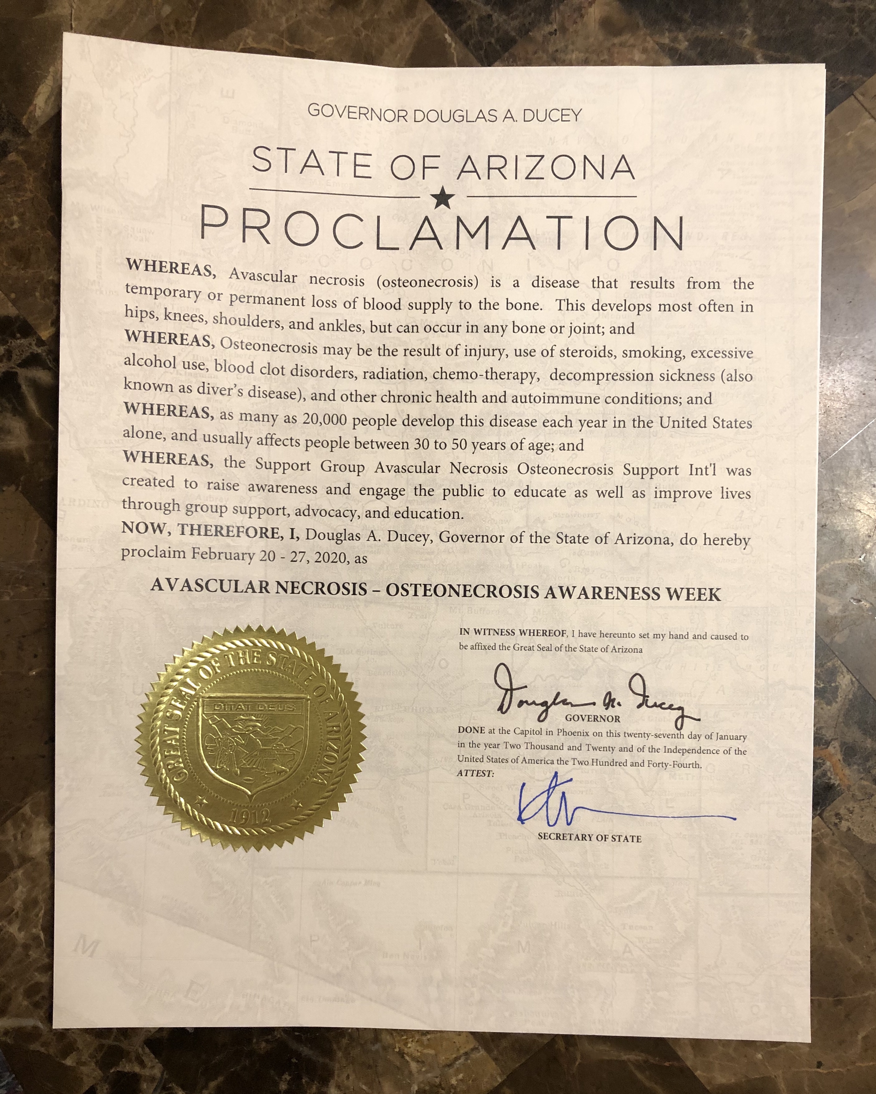
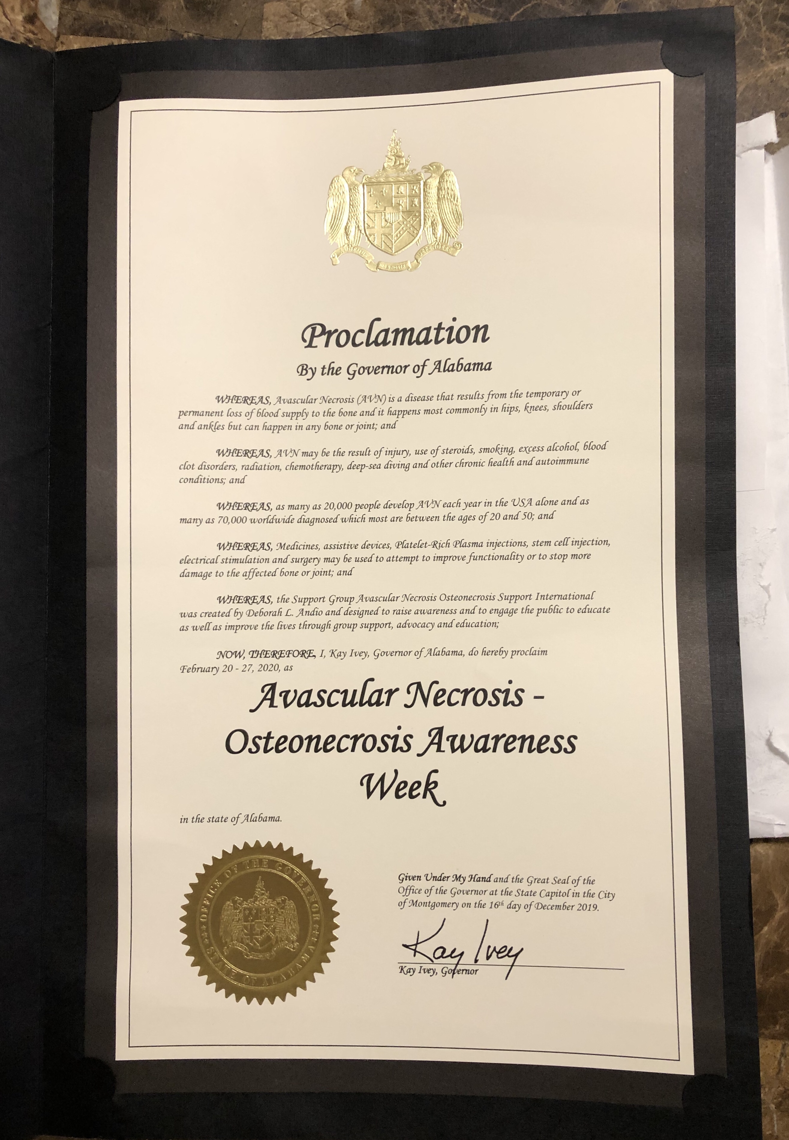
As we approach November 29, a day etched on our calendars as Avascular Necrosis-Osteonecrosis Awareness Day, the enthusiasm is palpable. Originating in states like Ohio, Pennsylvania, Iowa, Georgia, and Tennessee, and several other states this day has grown beyond its regional roots, capturing hearts and minds across the nation. A celebration of resilience, unity, and the strength of those battling this rare disease, the journey has just begun. We still have many states that have yet to recognize this awareness day.
In the spirit of reflection and progress, let’s take a closer look at what Avascular Necrosis-Osteonecrosis is and why it deserves the spotlight. Osteonecrosis occurs when the blood supply to a bone is disrupted, leading to the death of bone tissue. While it can affect anyone at any age, the rarity of this condition often results in delayed diagnosis and limited awareness.
Imagine a disease that doesn’t discriminate by age, gender, or background—a condition that can strike unexpectedly, altering lives in its wake. Avascular Necrosis-Osteonecrosis fits this description, emphasizing the importance of education and advocacy. It’s crucial to understand the pain that accompanies this condition. The affected bones, deprived of essential blood supply, lead to excruciating pain, impacting mobility and quality of life.
This awareness day isn’t just a commemoration; it’s a call to action. The commendable efforts of states like Ohio, Pennsylvania, Iowa, Georgia, and Tennessee have set the stage for a nationwide movement. Proclamations have been issued, voices have been amplified, and the journey towards recognition has begun.
As we celebrate the strides made, it’s essential to acknowledge the work that lies ahead. Advocacy is a journey, not a destination, and the goal is clear: to have all 50 states recognize and celebrate Avascular Necrosis-Osteonecrosis Awareness Day by 2024. This ambitious vision requires a collective effort.
To our esteemed congressmen, senators, and the President of the United States, we extend an invitation to join us in this noble cause. Avascular Necrosis-Osteonecrosis doesn’t discriminate based on political affiliations, and neither should our pursuit of awareness. We implore you to consider the impact of this rare disease on your constituents, urging you to champion the cause for an awareness day in every state.
It’s not a daunting task; it’s an opportunity for bipartisan collaboration, demonstrating a commitment to the health and well-being of the American people. A simple proclamation can go a long way in raising awareness, fostering empathy, and providing support to those affected by Avascular Necrosis-Osteonecrosis.
In the coming year, let’s collectively work towards making November 29 a day of national significance. Together, we can ensure that the stories of those touched by Avascular Necrosis-Osteonecrosis are heard, recognized, and celebrated from coast to coast. The journey has just begun, and with your support, we can light up the map with awareness, compassion, and unity.
Sincerely
Deb Andio
Founder
Avascular Necrosis-Osteonecrosis SuppooInt’l
http://www.avascularnecrosiseducation.com
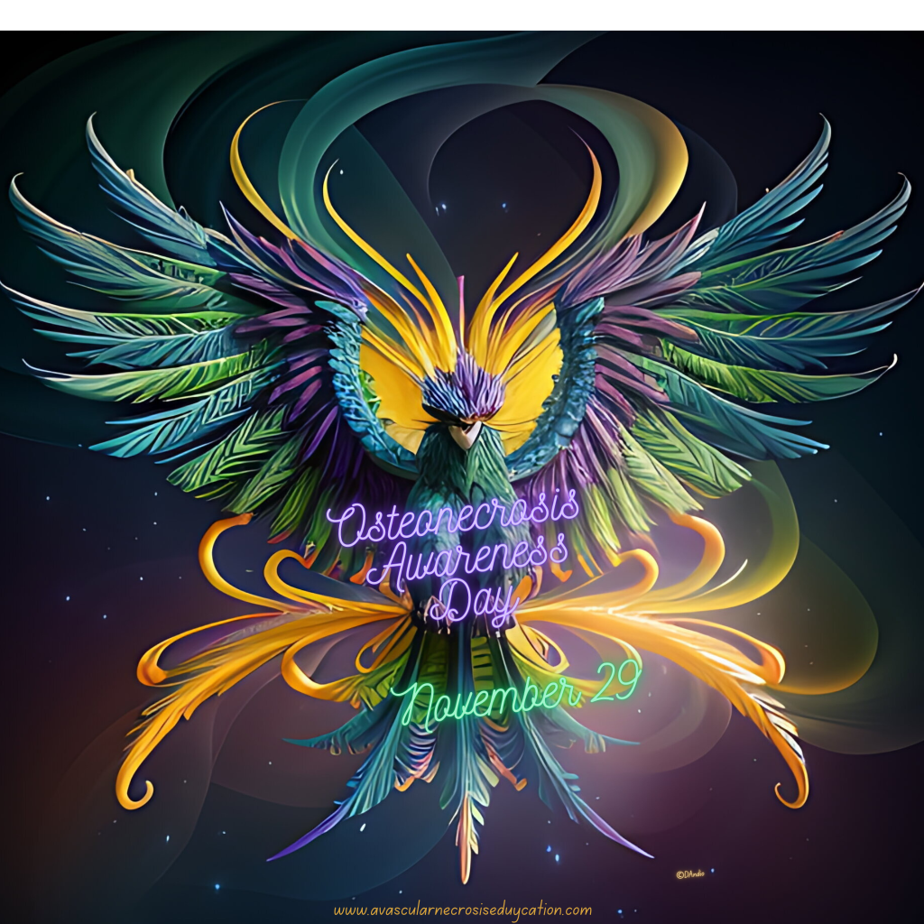
“Like the majestic Phoenix that rises from the ashes, those of us living with Osteonecrosis are on a journey of resilience and hope. As we continue to advocate for awareness, we believe that our Awareness Day will soon shine worldwide, illuminating the path for understanding, support, and compassion. Together, our voices will soar, echoing the strength that lies within each of us.”