Kummel Disease

Avascular necrosis (AVN) develops when a bone loses its blood supply. AVN goes by several other names, including Kummel disease, osteonecrosis, aseptic necrosis, and ischemic bone necrosis. AVN typically affects bone in the hips, thighs, knees and shoulders—although it can develop in any bone in the body.

Kummel disease is a rarely occurring variation of avascular necrosis that can affect the spine’s vertebrae, usually the thoracic spine (mid back) region. There are many different spine-related disorders that can contribute to disrupting a vertebral body’s blood supply, such as infection, osteoporosis, radiation therapy, steroid use, and metastatic spine tumors. Like other organs in the body, bone needs a healthy blood supply to rebuild itself, stay strong and support the spinal column.

Though Kummell disease is rare, researchers believe it’s becoming more prevalent as the aging population grows. People with osteoporosis and older adults are at a greater risk for developing this disorder.

Kummel Disease: A Not-So-Rare Complication of Osteoporotic Vertebral Compression Fractures

As our population ages, the prevalence of osteoporosis, its most common fragility fracture (vertebral compression fracture), and Kummel disease will increase.

Also if younger and you are diagnosed with osteoporosis your chances are higher to possibly develop Kummel Disease.


text ©Debla2017

Avascular Necrosis and Kummel Disease Share Some Similar Causes and Risk Factors

  • Injury: This is known as trauma-related avascular necrosis. A broken hip or vertebral fracture may lead to osteonecrosis.
  • Increased pressure within the bone: The pressure within the bone causes the blood vessels to narrow, making it hard for the vessels to deliver enough blood to the bone cells.
  • Certain risk factors: This is known as non-traumatic avascular necrosis. These risks include medical conditions and lifestyle choices that affect bone metabolism or bone remodeling.

AVN has several risk factors, including medical conditions and lifestyle choices that may increase your chances of developing the disorder:

  • Heavy alcohol use
  • Long-term corticosteroid use
  • Osteoporosis
  • Blood clots and arterial inflammation
  • Blood disorders (such as Sickle cell disease)
  • Radiation and chemotherapy
  • Pancreatitis
  • Gaucher disease (occurs when high amounts of fatty substances collect in the organs)
  • Decompression sickness (a condition causing gas bubbles in the blood)

How Doctors Diagnose Avascular Necrosis 

Diagnosing spinal AVN caused by trauma—also called Kummell disease—begins with a thorough review of your medical history and symptoms. This is all part of your physical exam.

After your physical exam, your doctor may order imaging scans to better see changes in your vertebrae.

Below are some of the tools your doctor may use to diagnose Kummell disease:

  • X-ray: This first-line imaging test can illuminate problems with your spinal bones. It’s not able to show early-stage bone problems, though.
  • Computed tomography (CT) scan: A CT scan provides a 3-dimensional picture of your bone. It also shows “slices” of bone, making the picture clearer than what x-rays and bone scans deliver.
  • Magnetic resonance imaging (MRI): This technology can produce very detailed images of your soft tissues and bones. The test is sensitive enough to see bone problems at their earliest onset, which x-ray is unable to do.
  • Bone scan: Also known as skeletal scintigraphy, bone scans are often used in people who have normal x-rays. A radioactive dye is injected into your affected bone and a picture of your bone is taken with a special camera. The picture shows how the dye travels through your bone and where normal bone formation is occurring.
  • Biopsy: A biopsy is a surgical procedure in which tissue from your affected bone is removed and studied. Although a biopsy is a conclusive way to diagnose AVN, it is rarely used because it requires surgery.
  • Functional evaluation of bone: Tests to measure the pressure inside a bone may be used when your doctor strongly suspects that you have AVN, despite normal x-ray, bone scan, and MRI results. These tests are very sensitive at detecting increased pressure within your bone, but they require surgery.

How Is Avascular Necrosis Treated?

The goals of treatment for AVN include improving your use of the affected joint, stopping further damage to the bone, and ensuring bone and joint survival.

If you have an early-stage form of Kummell disease, your doctor may prescribe medications to manage pain and/or inflammation. Physical therapy (PT) may be included in your treatment plan. A physical therapist can teach you how to exercise safely to protect your joints and bones while building strength, flexibility and endurance.

Though non-surgical treatments may help keep pain at bay, they are generally a temporary solution for people with Kummel disease. In many cases, spine surgery is necessary to prevent the condition from progressing to a point that it harms your quality of life.

Spine surgery for Kummel disease may have several purposes, depending on your specific symptoms. As such, your surgeon may use a single surgical approach or combine techniques to give you the best possible outcome. For example:

  • If you have developed abnormal kyphosis, your doctor may recommend osteotomy (surgical removal of bone) combined with spinal instrumentation and fusion. This combination of surgical procedures can reduce the size of the kyphotic curve, realign the spine and stabilize it.
  • Some patients with Kummel disease have pain and neurological symptoms, such as numbness or weakness. If symptoms are caused by nerve compression, a spinal decompression surgery (such as a foraminotomy) may be recommended to relief pressure on pinched nerves and prevent potentially permanent nerve damage.

Another surgical option for AVN is known as core decompression. This procedure involves removing the inner layer of bone, which reduces pressure within the bone, increases blood flow to the bone, and allows more blood vessels to form.

Core decompression works best in people who are in the earliest stages of avascular necrosis, often before the collapse of a joint. This procedure may reduce pain and slow progression of bone and joint destruction.

After core decompression, your surgeon may implant bone graft to help stimulate new bone growth and healing. Bone graft transplants healthy bone from a part of the body, such as the leg, to the diseased area. Several synthetic bone grafts are also available. Depending on the location and extent of the surgery, expect a lengthy recovery period, usually from 6 to 12 months.

As with all areas of medicine, researchers are continuously exploring treatments that may help people with AVN. One area of interest is therapies that increase the growth of new bone and blood vessels. These treatments have been used experimentally alone and in combination with other treatments, such as osteotomy and core decompression.

Your doctor will work with you to develop a custom treatment plan that addresses your symptoms and medical history.

Your Outlook with Avascular Necrosis (Kummel Disease)

For most people with avascular necrosis (also known as osteonecrosis, aseptic necrosis, and ischemic bone necrosis), treatment is an ongoing process. Your doctor may first recommend the least invasive approach and observe how you respond before progressing you to more substantial AVN therapies. If your condition affects your spinal vertebrae and was caused by trauma or injury (Kummell disease), several treatments may help prevent complications like spinal fracture, kyphosis deformity, and nerve pain from disrupting your life.

Educating On Bone Marrow Edema ©

Good Morning Pain Warriors Around The World
It is time to educate on the various causes of
Avascular Necrosis-Osteonecrosis
So twice a week we will post some educational info on a cause with links

Today Its Bone Marrow Edema

What is Bone Marrow Edema?
Bone marrow edema is a condition when excess fluids in the bone marrow build up and cause swelling. It is often caused by a response to an injury, such as a broken bone or a bruise, or a more chronic condition such as osteoporosis. Bone marrow edema most commonly occurs in the hips, knees and ankles. In this case, bone marrow edema of the knee is a main cause of localized knee and joint pain, and is only diagnosable via a Magnetic Resonance Imagining test (MRI).
It is usually caused by the following scenarios:
• Avascular necrosis, or “bone death”. This is when a small portion of the bone dies, and can result in a painful bone marrow edema
• Any type of knee bone trauma, including broken bones and bone bruises.
• Joint disorders such as osteoarthritis or osteoporosis. In this case, the knee joint is lacking the cushion that cartilage provides, which can lead to easier fracture and wear on the bone. Subsequently, if a fracture of the bone occurs, the injured area becomes susceptible to edema..
• Knee ligament injuries.
• A condition such as synovitis (an inflammation of the lining the joints, called synovial membranes).
• In rare conditions, bone tumor.
Symptoms of Bone Marrow Edema in the Knee
Bone marrow edemas may not bother you at all, or they may be painful and inconvenient. They can feel more intense than a muscular injury (for example, a muscle bruise) at times due to the nature of the bone. A muscle is capable of swelling, which increases blood flow to heal the area. Unfortunately, bones are not capable of swelling, and thus the fluid (edema) that collects in the marrow can create intense pressure within the bone, resulting in more intense pain. In fact, in many osteoarthritic patients, it isn’t the lack of cartilage that’s causing them pain, but rather the pressure due to the edema.

Some of the most common symptoms of bone marrow endema include:
• Varying degrees of pain, from mild to moderate, depending on the severity and Trauma.
• Swelling of the knee area.
• Inability to put full pressure on the knee to walk.
• Recurrent pain and tenderness.
• Bruising.
Treatment of Bone Marrow Edema in the Knee
Thankfully, most bone marrow edemas will settle down and heal on their own after the injury has subsided. For example, in some cases of osteonecrosis the bone will regenerate itself and heal the edema but note : not all cases of osteonecrosis or spontaneous osteonecrosis of the knee will have the ability to heal itself. Unfortunately, though, in the case of osteoarthritis, the edema may only get worse over time. In this circumstance, treatment options may be explored.
Traditional treatments for bone marrow edema usually involve rehabilitation through physiotherapy and rest. Ice, medications such as ibuprofen or acetaminophen, and even a crutch or a cane can help as well. There is one drug-facilitated treatment that uses a bisphosphonate and vitamin D mixture to help increase bone density. When this treatment is delivered via intravenous, it is found to be quite effective in reducing pain and increasing density. Other drugs that usually treat the vascular system have been found effective for bone marrow edema, in that they encourage blood flow and treat any vascular abnormalities that may exist in the bone and marrow.
In some more challenging cases, core decompression may be used. This is a type of surgery where a surgeon drills a hole into the affected part of the bone allowing that area of the bone to experience increased blood flow, form new blood vessels, and heal.
Another option is subchondroplasty, which can be especially effective for osteoarthritis patients. In this procedure, an x-ray determines where the edema is. The patient is then sedated, and a small needle injects a paste into the area of the edema. The paste then hardens and provides more strength and density to the bone. By improving the strength of the bone, it will enable the bone to deal with the pain of the edema and of the osteoarthritis.
http://louisvillebones.com/understanding-bone-marrow-edema/

Knee
https://www.g2orthopedics.com/bone-marrow-edema-in-the-knee/
https://www.researchgate.net/…/7224238_Bone_marrow_edema_in…
https://www.hindawi.com/journals/crirh/2018/7657982/

Hip Study
https://academic.oup.com/jcem/article/94/4/1068/2596208

https://www.ajronline.org/doi/10.2214/AJR.05.0086

https://www.ncbi.nlm.nih.gov/pubmed/15049532

https://www.ncbi.nlm.nih.gov/pmc/articles/PMC4972799/

Shoulder
https://www.sciencedirect.com/…/veterinary-sc…/osteonecrosis

Ankle
https://www.ncbi.nlm.nih.gov/pubmed/21189186
https://www.footanklesurgery-journal.com/…/S1268-7…/abstract


If you have #Osteonecrosis feel free to join our #group

AvascualrNecrosis/Osteonecrosis Support Int’l


©Debla2014

This slideshow requires JavaScript.

Joint Pain

 Congratulations !  If you’re like me you are one of the 30 million adults in the United States who suffer with joint pain, you know the pain often is debilitating. It can keep you from staying active and limits your mobility and it even makes daily chores seem impossible. What you might not know is that many doctors can treat joint pain with more than just pills or surgery. Beware though some doctors will tell you about one procedure and then change it once they know your insurance example Medicare I felt one doctor thought less of me as a patient because I am disabled so he changed or tried to change the procedure. It pissed me off because I was all set to get the procedure we discussed and then he changed it.

Both procedures were covered under Medicare so I felt betrayed as if I wasn’t good enough for the other injection.

Newsflash …..people on Medicare pay for the insurance they have. It’s not free , hell I pay more for insurance than when I was working.

But thankfully Medicare is good coverage. Low deductible.

It’s some doctors that treat you like a second class citizen.

Make sure this doesn’t happen to you.


Depending on the severity of your pain, injections can be another option for easing your joint pain and help to get you moving again.

Doctors use these injections to try to reduce inflammation and pain in your joints some come with side effects and some risks.

The injections range from corticosteroids, which have been around for decades, to newer ortho-biologic injections like platelet-rich plasma (PRP) , Stem Cell and placental tissue matrix (PTM)


You and your physician will decide which one is best based on your individual needs. The issue is finding doctors qualified to do these.

Not every injection is right for every patient, in my case I hate steroid injections, not only did it make my pain worse it also comes with the risk of developing osteonecrosis. Something I already have. I have noticed that it seems like doctors are quick to prescribe and inject steroids. I stand my ground and refuse. But that’s me.


So here are some facts to help you know more about the options.

Corticosteroid injections


Use: This injection is the first line of defense against osteoarthritis symptoms and other joint pain in shoulders, knees and hips. Corticosteroids can offer relief for two to three months, and reduce inflammatory cell activity in the joint. In some people.

Side effects and Risks : As with all injections, there’s a small chance of infection about one in 1,000 as well as Joint infection.

Nerve damage.

Thinning of skin and soft tissue around the injection site.

Temporary flare of pain and inflammation in the joint.

Tendon weakening or rupture.

Thinning of nearby bone (osteoporosis)

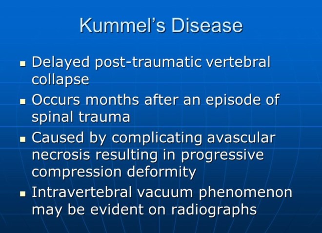
Osteonecrosis lack of blood supply to the bone

Raised blood sugar level

Whitening or lightening of the skin around the injection site

Cost: Most insurance covers the $100 -$200 usd cost of these injections. Your insurance provider may require that you try at least one corticosteroid injection first to see whether it works. If not, you may move on to a different therapy.


Hyaluronic acid injections

Use: Hyaluronic acid (HA) injections often are used when corticosteroid injections don’t work. But they usually are approved only for use in the knee.

In some instances, doctors consider an HA injection first if you don’t have obvious signs of inflammation. HA also is a better option if you have diabetes, as corticosteroids can raise blood sugar levels.

Also known as gel injections, HA injections are chemically similar to your natural joint fluid.

When you have osteoarthritis which is different than osteonecrosis lucky me I have both, the joint fluid becomes watery.

So, this injection helps to restore the fluid’s natural properties and works as a lubricant and a shock absorber.

HA is a cushion or a buffer against inflammatory cells in the joint.  In some cases, it can stimulate the knee to start producing more natural HA.” Some physicians also believe that HA helps reduce pain by coating nerve endings within the joint.

One treatment, which may consist of between one and three injections, usually offers symptom relief for four to five months, but sometimes up to one years. However, pain and stiffness will return. Most insurance companies only approve one HA injection every six months.

In knees with osteoarthritis, the joint fluid (called synovial fluid) can break down and not provide the cushioning your knee needs

Durolane

Euflexxa

Hyalgan

Orthovisc

Monovisc

Supartz

Synvisc, Synvisc-One

Depending on which type your doctor uses, you may get a single shot. Or you’ll get three to five injections spaced a week apart.


Side effects: There’s a 1-in-100 chance of an inflammatory reaction, The most common short-term side effects are minor pain at the injection site and minor buildup of joint fluid. These get better within a few days.


Cost: HA injections cost more — about $300 to $850 per injection, but most insurance companies cover the cost for knee injections.


Platelet-rich plasma (PRP) injections

Use: Platelet-rich plasma (PRP) injections can treat osteoarthritis joint pain, and are being thoroughly researched to understand their effects.

These injections use your own blood and platelets to promote healing. Platelets contain growth factors and proteins that aid healing in soft tissues. Research shows PRP injections can alter the immune response to help reduce inflammation,

Side effects: Side effects include a very low risk of infection and pain at the injection site. You must stop oral anti-inflammatory medications for a short amount of time if you get a PRP injection.

Cost: Insurance companies don’t generally cover PRP injections and you will pay between $400 and $1,300 per injection out-of-pocket.


Stem Cell Injections

The world’s most advanced regenerative injection treatments for treating knee pain due to arthritis, meniscus tears, traumatic ligament injuries, overuse conditions and other degenerative conditions.


Side effects : mild discomfort associated with the procedure. There is a very small risk of infection whenever aspirations and injections are performed. Nerve damage, vessel damage, and injury to other important structures are exceedingly rare


Placental tissue matrix (PTM) injections


Use: Placental Tissue Matrix (PTM) injections can very profoundly decrease the pain related to osteoarthritis.


These are injections of placental tissue, which is obtained after a healthy baby is delivered from a healthy mother. Research has discovered that there is a large number of growth factors in placental tissue that promote healing, Dr. Genin says.

Side effects: Side effects include a  low risk of infection and pain at the injection site. The placental tissue is “immune privileged,” which means the body would not have an adverse reaction to it.

Cost: Insurance companies don’t generally cover PTM injections; you will pay around $1,800 -$2500 per injection out-of-pocket.


Many of these injections often are effective in reducing or stopping your joint pain, but it’s important to remember that they may not keep the pain from returning, Dr. Schaefer says. In fact, they’re most effective when used with other therapies.


As a patient who has Osteonecrosis, Osteoarthritis, and other stuff I consider surgical options as a last resort only if other treatment options have failed. Unfortunately some treatments I cannot even afford to try. I wish the FDA would get a move on and approve some things so insurance companies can have this as a form of treatment.


Stem Cells


BONE MARROW AND FAT CELLS

The stem cells used in this point of care clinic are Autologous Cells that we take from your own body.  These cells are taken from your own Bone Marrow or Fat Cells.  The cells are your own Stem Cells and will not be rejected by your body.

Taking the Bone Marrow or Fat Cells from your body is relatively painless as a mild local anesthetic is used prior to harvesting.  These cells are processed to receive the most stem cell gain and then injected into the area of your body where you need the growth factors to go to work the quickest.  Your blood is also drawn and your platelet rich plasma is added to the Stem Cells taken from your Bone Marrow or Fat Cells to increase the activity of the growth factors.

It is important that these cells are used the day they are extracted from your body in order to insure they remain alive and active.  Our clinic does not grow extra stem cells from your Bone Marrow or Fat Cells to ensure that they are alive and active.  It is an FDA requirement that you receive your cells the same day they are harvested.

You get only the stem cells we extract from your body and there is no other manipulation used except extraction and preparation of the samples taken from your own body.  The cells are taken in a procedure that creates only mild discomfort or none at all.  Ninety nine percent of our patients experience no pain obtaining bone marrow or fat cells.

CORD STEM CELLS:

Embryo and Placenta stem cells can create certain types of cancers.  The cord blood Stem Cells should only be used if they are obtained from a healthy relative and you are a good match.  Cord Stem Cells that are used outside of the country or shipped to this country are illegal.  The FDA has found diseases in these grown cells and states that most of them are dead.  Even though the physicians supplying these Cord Cells claim they are safe to use, you should use extreme caution before considering these procedures.

ARE YOU A CANDIDATE FOR THESE STEM CELL PROCEDURES

REBUILDING JOINTS & SPINE: The Stem Cells that are obtained from your body are placed into all joints and spine to rebuild and regenerate new tissue growth as determined by the clinic physician.   There has been clinical evidence that new cartilage can be grown within your joint provided you are determined a candidate by the clinical physician.  Not all patients will be a candidate and may require joint replacement.

TORN TENDONS:  If the patients tendons are not completely torn this procedure will produce new tissue growth to regenerate torn tendons. Our clinic physician can only determine this with an initial visit and evaluation.

How Does PRP Therapy Work?

To prepare PRP, a small amount of blood is taken from the patient. The blood is then placed in a centrifuge. The centrifuge spins and through a multi-functional process separates the plasma from the blood producing the PRP. This increases the concentration of platelets and growth factors up to 500% also increasing hMSC (human stem cells) proliferation as a function of 8-day exposure to platelet released concentrations 10x. (x= increase above native levels)

When PRP is injected into the damaged area it stimulates the tendon or ligament causing mild inflammation that triggers the healing cascade. As a result new collagen begins to develop. As this collagen matures it begins to shrink causing the tightening and strengthening of the tendons and ligaments of the damaged area.

What is Platelet Rich Plasma?

Platelet Rich Plasma or PRP is blood plasma with concentrated platelets. The concentrated platelets found in PRP include growth factors among the huge reservoirs of bioactive proteins that are vital to initiate and accelerate tissue repair and regeneration. These bioactive proteins increase stem cell production to initiate connective tissue healing, bone regeneration and repair, promote development of new blood vessels and stimulate the wound healing process.

PRP Regenerates Tendons & Ligaments

Tendons connect the muscle to the bone making it possible for you to do many everyday physical activities. Overuse or damage to the tendon over a long period of time causes the collagen fibers in the tendons to form small tears, a condition called tendonitis. Damage to tendons most often occurs in the knees, ankles, hips, spine, elbows, shoulders, and wrists.

Ligaments are composed of collagen fibers that hold one bone to another, stabilizing the joint and controlling the range of motion. When a ligament is damaged, it is no longer able to support the bones in the joint, which often leads to pain symptoms. The instability causing the pain in your joints does not always show up on high tech imaging equipment. Through a thorough neurological and orthopedic evaluation Dr. Baum can determine which ligaments and tendons are unstable due to injury, wear or tear.

Tendons and ligaments have a poor blood supply and they do not usually heal from damage. Combined with the stress of day-to-day activities tendons and ligaments become inefficient causing degeneration of the joint which leads to chronic pain and weakness. Patients who experience chronic pain may not even remember when the injury occurred.

How Does PRP Compare With Cortisone Shots?

Studies have shown that cortisone injections may actually weaken tissue. Cortisone shots may provide temporary relief and stop inflammation, but may not provide long term healing. PRP therapy is healing and strengthening these tendons and ligaments and in some cases thickening the tissue up to 40%.

Treatment Plan

PRP injections with guided ultrasound can be performed on tendons and ligaments all over the body. Cervical, thoracic and lumbar spine, degenerative disc disease, arthritic joints shoulder pain, hip pain, and knee pain, even the smaller joints of the body can all be treated effectively with PRP. Dr. Baum will determine whether prolo solution, Platelet Rich Plasma or a combination of both will be the most effective form of treatment for you during his initial consult and evaluation.

Frequency Of Treatments

While responses to treatment vary, most people will require 3 to 6 sets of injections of PRP. Each set of treatments is spaced 4 to 6 weeks apart.

Is PRP Right For Me?

If you have degenerative spine or joint disease, a tendon or ligament injury, laxity or tear and traditional methods have not provided relief then PRP therapy may be the solution. It will heal tissue with minimal or no scarring and alleviates further degeneration and builds new tissues. There will be an initial evaluation with Dr. Baum to see if PRP therapy is right for you.

What Can Be Treated?

Platelet Rich Plasma injections helps regenerate all areas of the body including the cervical, thoracic and lumbar spine, wrists, elbows, shoulders, hips, knees and ankles as well as tendons and ligaments all over the body.  Dr. Baum is one of the few physicians performing PRP procedures to all areas of the spine.  Our clinic treats patients with sports injuries, arthritic and degenerative joints and degenerative disc disease. More specific injuries including tennis elbow, carpal tunnel syndrome, scoliosis, ACL tears, shin splints, rotator cuff tears, plantar faciitis and iliotibial band syndrome may all be effectively treated with PRP.

What Are The Potential Benefits?

Patients can see a significant improvement in symptoms as well as a remarkable return of function. This may eliminate the need for more aggressive treatments such as long-term medication or surgery.

Special Instructions

You are restricted from the use of non-steroid anti-inflammatory medications (NSAIDs) one week prior to the procedure and throughout the course of treatments.

Initially the procedure may cause some localized soreness and discomfort. Most patients only require some extra-strength Tylenol to help with the pain. Ice and heat may be applied to the area as needed.

How Soon Can I Go Back Regular Activities?

PRP therapy helps regenerate tendons and ligaments but it is not a quick fix. This therapy is stimulating the growth of new tissue requiring time and rehabilitation. Under Dr. Baum’s supervision patients will begin an exercise program immediately following the first procedure. During the treatment program most people are able to resume normal activities and exercise.

Platelet Rich Plasma (PRP) Matrix Graft by David Crane, MD and Peter A.M. Everts PhD

PRP application techniques in musculoskeletal medicine utilize the concentrated healing components of a patient’s own blood—reintroduced into a specific site—to regenerate tissue and speed the healing process

PRP INJECTION APPLICATION SITES

Spine

Cervical/Thoracic/Lumbar/Sacral

Shoulders & Elbows

Wrist & Hand

Hip/Pelvis

Knee & Lower Leg

Ankle & Foot

Fingers & Toes

Arthritic Joints

Osteoarthritis

Some Osteonecrosis


Information

http://www.prolotherapy.com/PPM_JanFeb2008_Crane_PRP.pdf

https://drjamesbaum.com/wp-content/uploads/stemcells2002-0109.pdf


Important Videos Everyone Should watch on Biologics

https://drjamesbaum.com/2013/07/the-science-of-mesenchymal-stem-cells-and-regenerative-medicine/


Scientific Papers on Research of Stem Cells

https://drjamesbaum.com/stem-cells/scientific-papers/

knnz

I will be posting this in my other blog section also

Tired of Doctors? I am also.

If you are like me,your sick of seeing doctors , some see so many my heart breaks for them, pcp, orthos, hematologist, cardiologists, pain management, endocrinologist dentists, optometrist whatever it just gets tiring. then you might get so nervous at your appointment you forget to ask specific questions.

Or maybe your anxiety has gotten you to a place where your heart is beating out of your chest, and your voice quivers or you just become blank.

Either way, having a list of things you mean to ask will help not only you but your doctor be able to better communicate.

So here we go:

1. What is the exact name of whatever is wrong with me? 1a if Osteonecrosis what stage is it? 1-2-3-4-5-5a-6

2. Is this something that is treatable and may possibly go away, or heal on its own or is this something that is chronic?

3. What are the short-term and long-term prognoses?

4. What are the short-term and long-term goals with treatment?

Example : Prp injections- physical therapy- hyperbaric oxygen-water therapy- stem cell treatments- joint replacement .

5. Is this something that genetically I can pass down to my child(ren)?

5a. Will you check me for underlying clot issue like Factor V Leiden-MTHFR-eNOS

6. What do you recommend as far as treatment?

7. What are the medications you recommend? I heard and read statins like Zocor and Lipitor can help lower lipids (fat) in the blood which can help blood flow better. I personally took simvastatin for 6 m and it helped me. I also during that time changed how I ate to consume less fatty meat cheeses etc

To lower my cholesterol on my own and I must say I am grateful I tried this.

Not only do I love eating REAL food but my blood work shows fantastic results and my pain is less.

So it’s good to talk to your doctor about this stuff also. Of course your doctor is not a specialist in nutrition which is kinda sad , you’d think they would teach that in medical school. But a good plant based diet was a great change for me.


8. How will this condition and/or medications affect my life?

9. Is there a specialist I can see? So I may try to avoid a joint replacement?

10. Will this condition or medications affect my ability to exercise? Walk-Bike- Swim.

Will this be temporary?

11. Is there a special diet that might ease symptoms or improve this condition?

For me personally it is plant based. I will eat meat (beef) rarely . Chicken or fish 1x a week but I am all about plant based.

My pain is lessened by over 60percent. I still have pain I’m not cured but I’m not in constant misery anymore. I rarely take a pain pill.

Never start or stop anything without talking to your doctor.

As always, if you have any concerns about your health, it’s always best to consult your primary care physician.

~~~~~~~~~~~~~~~~~~~~~~~~~~~~~~~~~~~~~~~~~~~~~~~~~~~~~~~~~~~~~~~~~~~~~~~~~~~~~~


Specific Questions To Ask Your Otho

What stage is osteonecrosis?
Will I be checked to make sure it also is not affecting other joints since I have pain in other bones/joints?
What is your treatment plan for Osteonecrosis ?
Why is this procedure being recommended? Are there alternatives?
What are the benefits of this procedure in terms of pain relief, functioning/mobility? How long will the benefit last?
What are the risks involved?
What is the success rate for this procedure?
What is the procedure called? How is it done?
How many patients with osteonecrosis do you see a year?
If Surgery Will this surgery solve the problem? Will any more surgery be required in the future?
How many of these procedures are annually performed at this hospital?
How many patients with osteonecrosis do you see a year?
What percentage of patients improve following the procedure?
What will happen if I don’t have the surgery now?
If I want a second opinion, whom can I consult?
Will I need any tests or medical evaluations prior to the surgery?
What kind of anesthesia will be used?
Are there possible after effects or risks?
Will I meet with the anesthesiologist in advance? Will her or she know my needs/allergies?
What kind of implant or prosthesis will be used? What are the outcomes using this device?
How long will it last?
Will I have pain following the procedure? What pain relief or pain control measures will I be given?
How long will the recovery take? What are my limitations during recovery?
Will I need assistance at home afterwards? For how long?
What will discharge instructions be?
Will I have any disability following surgery? Will I need physical therapy?
When can I return to work? When can I drive my car? When can I have sexual activity?
Are there any materials about this surgery that I can review?
What will I have for pain management?
Are their any patients I can ask about their experience?


Here is some info you may be interested in.

Clinical Trials

Atorvastatin to Prevent Avascular Necrosis of Bone in Steroid Treated Exacerbated Systemic Lupus Erythematosus

https://clinicaltrials.gov/ct2/show/NCT00412841

Statin therapy decreases the risk of osteonecrosis in patients receiving steroids.

https://www.ncbi.nlm.nih.gov/m/pubmed/11347831/

Aseptic osteonecrosis of the hip in the adult: current evidence on conservative treatment

https://www.ncbi.nlm.nih.gov/pmc/articles/PMC4832407/

Steinberg Stages Of Osteonecrosis-Avascular Necrosis

Steinberg Staging Of Avascular Necrosis/ Osteonecrosis

History and etymology

It is based on the radiographic appearance and location of lesion. It primarily differs from the other systems by quantifying the involvement of femoral head which allows direct comparison between series1. Seven stages of involvement are identified. Following staging, extent of involvement of femoral head is recorded as mild, moderate or severe.

Classification

stage 0: normal or non-diagnostic radiographs, MRI and bone scan of at risk hip (often contralateral hip involved, or patient has risk factors and hip pain)

stage I: normal radiograph, abnormal bone scan and/or MRI

stage II: cystic and sclerotic radiographic changes

stage III: subchondral lucency or crescent sign

stage IV: flattening of femoral head, with depression graded into

mild: <2 mm

moderate: 2-4 mm

severe: >4 mm

stage V: joint space narrowing with or without acetabular involvement

stage VI: advanced degenerative changes

Quantification of extent of involvement is necessary for stages I to V:

stage I and II

A, mild: <15% head involvement as seen on radiograph or MRI

B, moderate: 15% to 30%

C, severe: >30%

stage III

A, mild: subchondral collapse (crescent) beneath <15% of articular surface

B, moderate: crescent beneath 15% to 30%

C, severe: crescent beneath >30%

stage IV

A, mild: <15% of surface has collapsed and depression is <2mm

B, moderate: 15% to 30% collapsed or 2 to 4mm depression

C, severe: >30% collapsed or >4mm depression

stage V

A, B or C: average of femoral head involvement, as determined in stage IV, and estimated acetabular involvement.

Steinberg Staging Osteonecrosis

See also

Avascular Necrosis Of The Hip

Fixated and Arlet Staging

Legg-Calvé Perthes Disease

AVN CharityUK

References

1. Steinberg ME, Hayken GD, Steinberg DR. A quantitative system for staging avascular necrosis. J Bone Joint Surg Br. 1995;77 (1): 34-41. Pubmed citation

How Our Joints Work and Move

How Our Joints Work

Joints are the place where two or more bones meet. All of your bones, except for one (the hyoid bone in your neck), form a joint with another bone. Joints hold your bones together and allow your rigid skeleton to move.

Fixed joints
Some of your joints, like those in you skull, are fixed and don’t allow any movement. The bones in your skull are held together with fibrous connective tissue.

Slightly movable joints
Other joints such as those between you vertebrae in your spine, which are connected to each other by pads of cartilage, can only move a small amount.

Synovial movable joints
Most of your joints are “synovial joints”. They are moveable joints containing a lubricating liquid called synovial fluid. Synovial joints are predominant in your limbs where mobility is important. Ligaments help provide their stability and muscles contract to produced the desired movement. The most common synovial joints are listed here:

Ball and socket joints, like your hip and shoulder joints, are the most mobile of joints in the human body. They allow you to swing your arms and legs in many different directions.
Hinge joints like in your knee and elbow , enable movement similar to opening and closing a hinged door.
The pivot joints in your neck allow you to turn you head from side to side.
Ellipsoidal joints such as the joint at the base of your index finger, allow bending and extending, rocking from side to side, but rotation is limited.
Gliding joints occur between the surfaces of two flat bones that are held together by ligaments. Some of the bones in your wrists and ankles work by gliding against each other.

Conyloid joints. These joints are similar to ball-and-socket joints, just without the socket (the “ball” simply rests against another bone end).
Saddle joints are found in your thumbs, the bones in a saddle joint can rock back and forth and from side to side, but they have limited rotation.


yoga

Avascular Necrosis – Osteonecrosis eBooklet © ™️®️


Click link below


AVN Awareness Booklet    

To access link on cell press on above link and you will be re-directed to Booklet.

If on computer just click above link.

🌻 Hi, I’m Deborah Andio

I wrote this booklet in 2016  to help patients like myself and their families understand avascular necrosis- osteonecrosis  and give helpful ideas to patients and help our doctors understand the pain we often feel.

My goal is throughout this booklet is to first let you know you are not alone .

Those who also have been diagnosed with avn know exactly how your feeling,scared, afraid, searching for knowledge and resources and coming up with very little.

That’s why I started a support group and wrote this booklet.

Disclaimer

  • This booklet is not intended as a substitute for the medical advice of physicians. The reader should regularly consult a physician in matters relating to his/her health and particularly with respect to any symptoms that may require diagnosis or medical attention.
  • No part of this eBook may be reproduced or transmitted in any form or by any means, electronic or mechanical, including photocopying, recording or by any information storage and retrieval system, without written permission from the author.
  • The information provided within this eBook is for general informational purposes only.
  • Copyright © protected
  • While I try to keep the information up-to-date and correct, there are no representations or warranties, express or implied, about the completeness, accuracy, reliability, suitability or availability with respect to the information, products, services, or related graphics contained in this eBook for any purpose. Any use of this information is at your own risk.

I give an actual copy to members of my support group. But many felt the information was so helpful, I wanted to share it with everyone.

I have recently completed a better spell check and next print will have all errors corrected.

No-one can use any contents of this booket words or graphics

Also booklet will be updated every 2 years

2014 check

2016

2018

2020

I am continually writing to our Senators state by state asking for an awareness day proclamation.

So AVN-ON can get the recognition it deserves.


Supprt Group Link  Avascular Necrosis/ Osteonecrosis Support Int’l

Wishing you all a pain free and relaxingI am

God Bless

Debbie


ChronicallyGratefulDebla2018© ™️

Debla©2014-2019 ™️


All rights reserved. Thia booklet on line or in print may not be reproduced in any form, stored in any retrieval system, or transmitted in any form by any means—electronic, mechanical, photocopy, recording, or otherwise—without prior written permission of the publisher, except as provided by United States of America copyright law. For permission requests, write to the author on this web site.

©Debla2014


computer